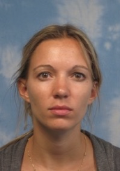
Welcome Frédéric Murisier!

Hello! My name is Frédéric Murisier, and I am excited to join Professor Jean-Yves Roignant’s team as a PhD student. I am eager to contribute to the lab’s research on RNA methylation as part of my doctoral journey.
I am originally from Switzerland. I completed my Bachelor’s degree in Biomedical Sciences and my Master’s degree in Bioinformatics at the Université Libre de Bruxelles in Belgium. Before joining this team, I worked for over two years in an epigenetics lab focused on cancer research, where I developed a strong foundation in computational biology and genomics.
My goal is to pursue a PhD to deepen my knowledge and further develop my skills in bioinformatics, particularly in the context of gene regulation mechanisms. I am passionate about unraveling complex biological systems through data analysis and am eager to expand my expertise in this exciting field.
Outside of research, I enjoy climbing and running, always looking for new challenges and ways to push my limits.
I am thrilled to begin this new adventure and look forward to collaborating with everyone in the lab!
IDEAS COMPETITION 2025 10 years – 10 ideas – 100 possibilities to carry on the spark
What should our society look like in 2035?
https://www.franxini.ch/en/idee
Which impulses do we need today to shape the Switzerland of tomorrow?
Do you have an idea?
We are looking for sparks that will create a brighter future!
2035 has established itself as the target year for many ambitious global projects – but where do we want to be as Switzerland in 10 years’ time? And how can we achieve this?
In 2025, the Reatch ideas competition is coming back with a new, exciting format for the final, which is all about the next 10 years ahead: we are looking for ideas and projects that generate a momentum for the future.
Let’s bring society, politics and science together to shape the Switzerland of tomorrow!
Deadline: March 12, 2025
Two Faculty positions: Aarhus University in Denmark (deadline April 2025)
Biochem Soc Trans, auth: group Vastenhouw
The role of transcription bodies in gene expression: what embryos teach us
Martino Ugolini 1 , Nadine L Vastenhouw 1
Affiliations
- PMID: 39912709
- DOI: 10.1042/BST20240599
Abstract
Transcription does not occur diffusely throughout the nucleus but is concentrated in specific areas. Areas of accumulated transcriptional machinery have been called clusters, hubs, or condensates, while transcriptionally active areas have been referred to as transcription factories or transcription bodies. Despite the widespread occurrence of transcription bodies, it has been difficult to study their assembly, function, and effect on gene expression. This review highlights the advantages of developmental model systems such as zebrafish and fruit fly embryos, in addressing these questions. We focus on three important discoveries that were made in embryos. (i) It had previously been suggested that, in transcription bodies, the different steps of the transcription process are organized in space. We explore how work in embryos has revealed that they can also be organized in time. In this case, transcription bodies mature from transcription factor clusters to elongating transcription bodies. This type of organization has important implications for transcription body function. (ii) The relevance of clustering for in vivo gene regulation has benefited greatly from studies in embryos. We discuss examples in which transcription bodies regulate developmental gene expression by compensating for low transcription factor concentrations and low-affinity enhancers. Finally, (iii) while accumulations of transcriptional machinery can facilitate transcription locally, work in embryos showed that transcription bodies can also sequester the transcriptional machinery, modulating the availability for activity at other sites. In brief, the reviewed literature highlights the properties of developmental model organisms that make them powerful systems for uncovering the form and function of transcription bodies.
Keywords: Developmental model systems; Gene regulation; Nuclear organization; Sequestration; Transcription bodies.
Nature, co-auth: J. Larsch
Transcriptomic neuron types vary topographically in function and morphology
Inbal Shainer # 1 2 , Johannes M Kappel # 1 3 , Eva Laurell 1 , Joseph C Donovan 1 , Martin W Schneider 1 , Enrico Kuehn 1 , Irene Arnold-Ammer 1 , Manuel Stemmer 1 , Johannes Larsch 1 4 , Herwig Baier 5
Affiliations
- PMID: 39939759
- PMCID: PMC11864986
- DOI: 10.1038/s41586-024-08518-2
Abstract
Neuronal phenotypic traits such as morphology, connectivity and function are dictated, to a large extent, by a specific combination of differentially expressed genes. Clusters of neurons in transcriptomic space correspond to distinct cell types and in some cases-for example, Caenorhabditis elegans neurons1 and retinal ganglion cells2-4-have been shown to share morphology and function. The zebrafish optic tectum is composed of a spatial array of neurons that transforms visual inputs into motor outputs. Although the visuotopic map is continuous, subregions of the tectum are functionally specialized5,6. Here, to uncover the cell-type architecture of the tectum, we transcriptionally profiled its neurons, revealing more than 60 cell types that are organized in distinct anatomical layers. We measured the visual responses of thousands of tectal neurons by two-photon calcium imaging and matched them with their transcriptional profiles. Furthermore, we characterized the morphologies of transcriptionally identified neurons using specific transgenic lines. Notably, we found that neurons that are transcriptionally similar can diverge in shape, connectivity and visual responses. Incorporating the spatial coordinates of neurons within the tectal volume revealed functionally and morphologically defined anatomical subclusters within individual transcriptomic clusters. Our findings demonstrate that extrinsic, position-dependent factors expand the phenotypic repertoire of genetically similar neurons.